Sebastian Knust¹, Andreas Meyer², André Spiering¹, Christoph Pelargus¹, Andy Sischka¹, Peter Reimann², and Dario Anselmetti¹
¹ Experimental Biophysics and Applied Nanoscience, Faculty of Physics, Bielefeld University
² Theoretical Physics and Soft Matter Theory, Faculty of Physics, Bielefeld University
Abstract
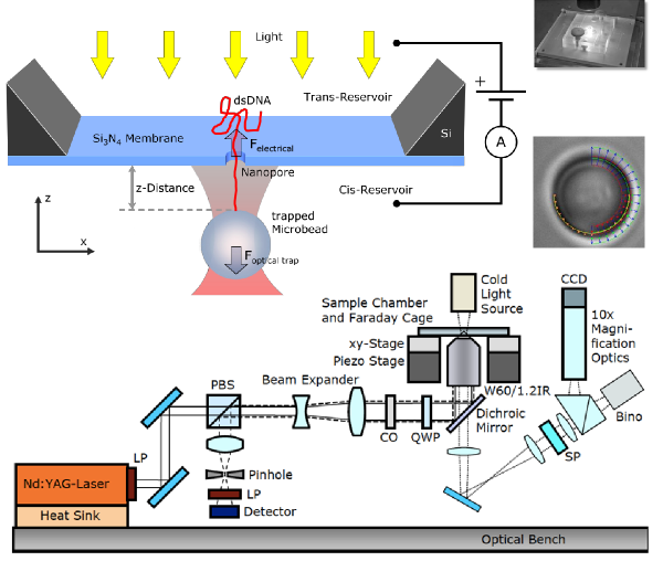
We measured forces acting on DNA during translocation through a nanopore with Optical Tweezers. A video-based force detection and analysis system was developed, allowing for virtually interference-free axial force measurements with an overall force resolution of ±0.5 pN at a sample rate of 123 Hz [1].
We previously measured the translocation of dsDNA through 20 nm thick Si3N4 membranes (0.1 pN/mV for pores ≥ 30 nm) [2, 3]. Lipid coating as well as carbon nanomembranes and graphene with a thickness of 3 nm and 0.3 nm respectively allow for even more sensitive measurements.
Experimental Setup
- Video-based force detection → reduced interference
- Calibration via Stokes friction and Allan variance
- Force sensitivity better than 0.5 pN at 123 Hz sample rate
Translocation Theory
- Dynamics dominated by electrohydrodynamic effects (electro-osmotic flow)
- Modeling with combination of Poisson, Nernst-Planck and Stokes equations
- Mere zero surface charge on coated membrane does not explain high forces
- → Introduction of slip length at the DNA-solution-interface
- Supported by theoretical treatment of DNA nanostructure [3] and recent MD simulations [4]
Results:
Slip length \(\)
Surface charge for Si3N4 \(\)
Nanomembrane Preparation
Graphene
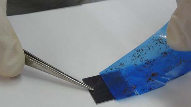
- Mechanical exfoliation with nitto tape
- Automated searching routine for graphene flakes
- Transfer on chip with cellulose polymer
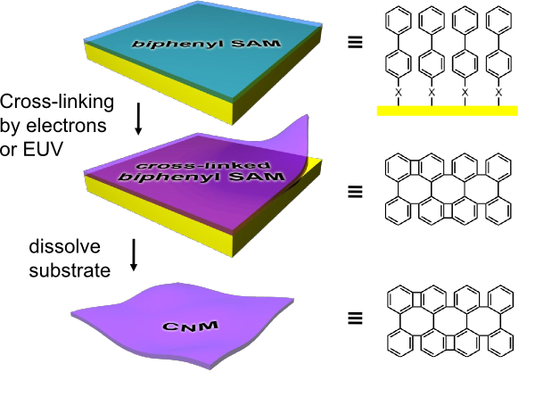
Carbon nanomembranes
Resulting thickness of 1-3 nm
Nanopore Preparation
- Zeiss Orion Plus HIM
- 0.35 nm imaging resolution
- Pore sizes as small as 5-6 nm
Graphene
Strongly localised heating phenomena (plasmon?)
→ Melts polystyrene beads
→ Dissipates biotin-streptavidin bond between bead and DNA
Carbon Nanomembranes
- 3 nm membrane thickness
- 70 nm NP size
- 50 mV
- 20 mM KCl
- Interference caused by silicon chip geometry
References
[1] S. Knust et al., Video-based and interference-free axial force detection and analysis for optical tweezers. Rev. Sci. Instr. 83, 103704 (2012)
[2] A. Spiering et al., Nanopore translocation dynamics of a single DNA-bound protein. Nano Lett. 11, 2978 (2011)
[3] A. Sischka et al., in preparation
[4] S. Kesselheim, W. Müller, C. Holm, Origin of Current Blockades in Nanopore Translocation Experiments. Phys. Rev. Lett. 112, 018101 (2014)
Downloads / Links
- PDF of poster
- We are selling Optical Tweezers! Visit us at picotweezers.com


